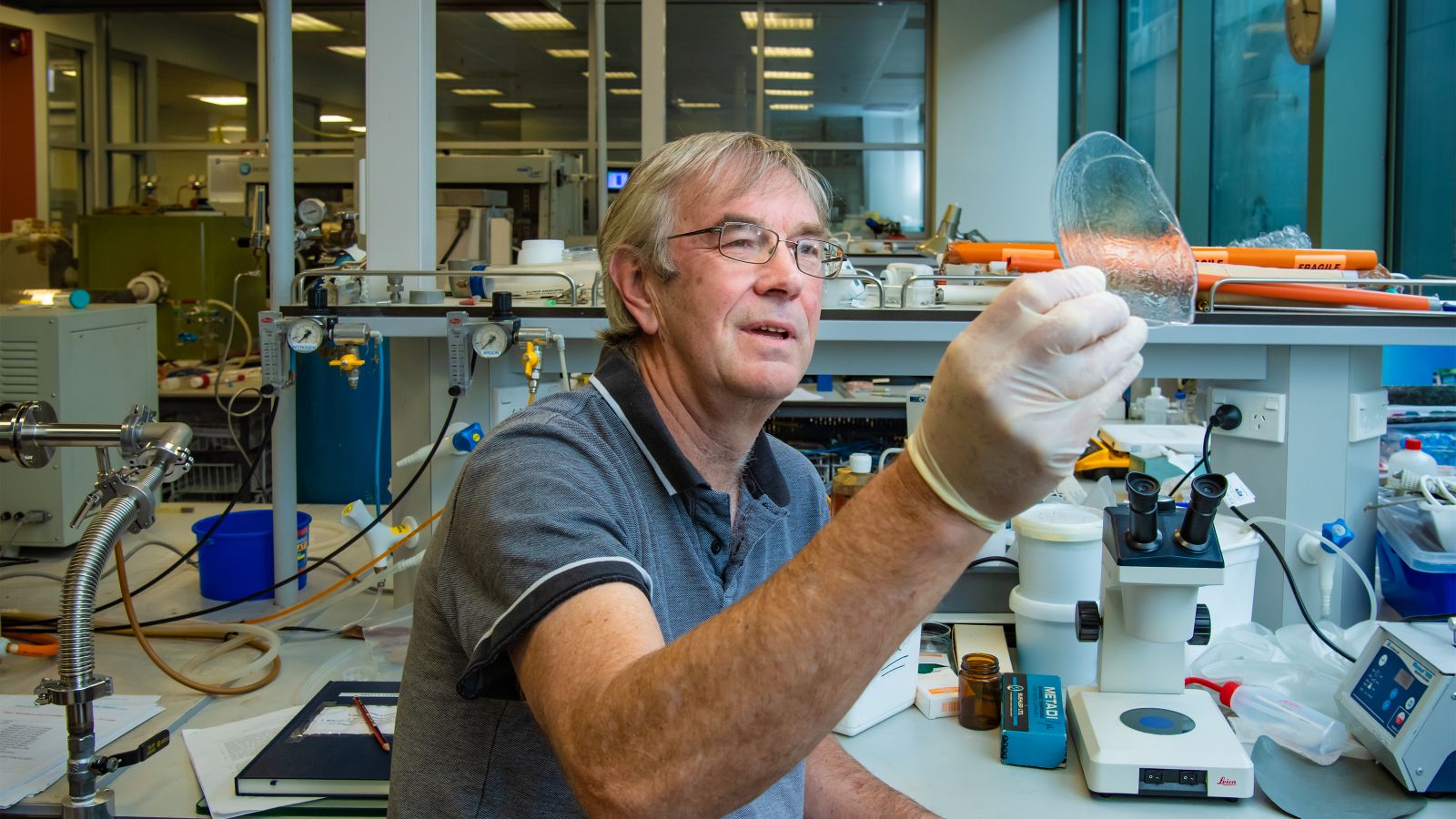
Associate Professor Andy Edgar from the University’s School of Chemical and Physical Sciences participated in a recent research project with the University of Saskatchewan in Canada to develop new radiation treatments for inoperable tumours, particularly in the brain or spine.
This research project was investigating microbeam radiation therapy, which uses high-powered beams of radiation no wider than a human hair to target tumours while leaving the healthy tissue around them untouched.
The University of Saskatchewan researchers successfully identified the ideal strength of these beams to target the cancer. They also developed a high-dose radiation detector to ensure the dose of radiation is precise and controlled—which is where Associate Professor Edgar comes in.
The high-dose detector uses glass containing a rare Earth ion called Samarium that helps measure the radiation doses. Samarium in the glass glows orange when exposed to a blue laser before x-ray irradiation, and red after exposure. By creating an image of the red and orange areas and comparing the different amounts of red and orange the researchers could determine the level of radiation being delivered during the microbeam radiation therapy.
The glass was created by Associate Professor Edgar in the University’s laboratories in Wellington. Associate Professor Edgar, who recently retired, has collaborated with the team from the University of Saskatchewan for many years after meeting the lead researcher at a conference 12 years ago. The collaboration included spending time at the University of Saskatchewan working on this project directly and hosting Canadian collaborators at Victoria University of Wellington.
“I came up with the idea to use this particular family of glasses in the project, as well as designing and creating the final glass composition used in the detector,” Associate Professor Edgar says. “It took three different combinations until we found the right combination for the glass.
Creating the glass is a complex process.
“It’s not like creating window glass,” Associate Professor Edgar says. “These specialised glasses need to be made in an inert atmosphere. 30 thirty seconds to pour red-hot glass at 1000 degrees Celsius and sandwich it between two metal plates which are themselves at 400 degrees Celsius—it’s certainly exciting but somewhat stressful.”
Before shipping the glass to Canada, Associate Professor Edgar tested it here. In both his tests and the tests completed in Canada, he was excited to see that the glass could produce very detailed images of the radiation, detecting wires only 1/25th of a millimetre wide.
“If you’ve ever seen an x-ray image, the edges of structures can be blurry because of the way x-rays are traditionally detected,” Associate Professor Edgar says. “The glass we have made can detect very fine and much sharper detail.”
This research project could provide quite a dramatic development in cancer treatment.
“With this understanding of the beam strength required and the ability to measure it, medical professionals can deliver much higher targeted doses of radiation to patients,” he says. “Current, more imprecise methods of delivering radiation therapy must be limited, because they damage healthy tissue as well as cancerous tissue. This method could be used to safely deliver stronger doses to patients compared to current treatments.”
Associate Professor Edgar also says any application that requires imaging with this level of detail would benefit from this discovery.
The work on glasses formed part of long-term Ministry of Business, Innovation, and Employment funded programme on materials for radiation imaging and detection by Associate Professor Edgar and Professor Grant Williams, also from Victoria University of Wellington. Before retiring in late 2019, Associate Professor Edgar's last contribution to the programme was the development of two systems for portable x-ray imaging.
